Standard Layer
When used for Epi-Illumination
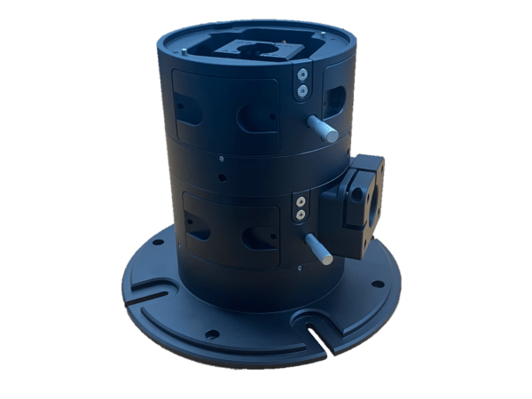
On the openFrame the Single Epi Layer provides a means of coupling a single illumination source to the microscope. This is the preferred solution for applications that only require a single illumination source. It is also the recommended solution for multi-modal illumination, for example when combining widefield fluorescence imaging with simultaneous photostimulation. In such cases, the second illumination source would mount on a Single or Dual Epi Layer in the Flexi Layer position (see below).
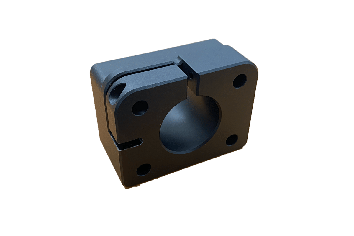
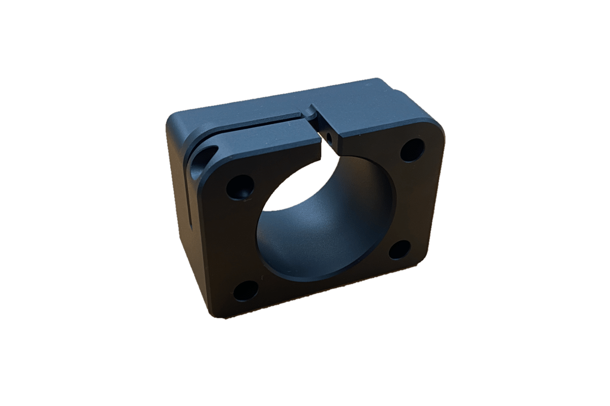
When using the Single Epi Layer, there are two possible mounting points located on opposite sides of the module. While only one of these is used at a time, the two positions provide more flexibility and avoid spatial conflicts with components on other layers when bulky illuminators are used. It also allows a custom illuminator to be built in parallel without decommissioning the microscope from it's day-to-day use
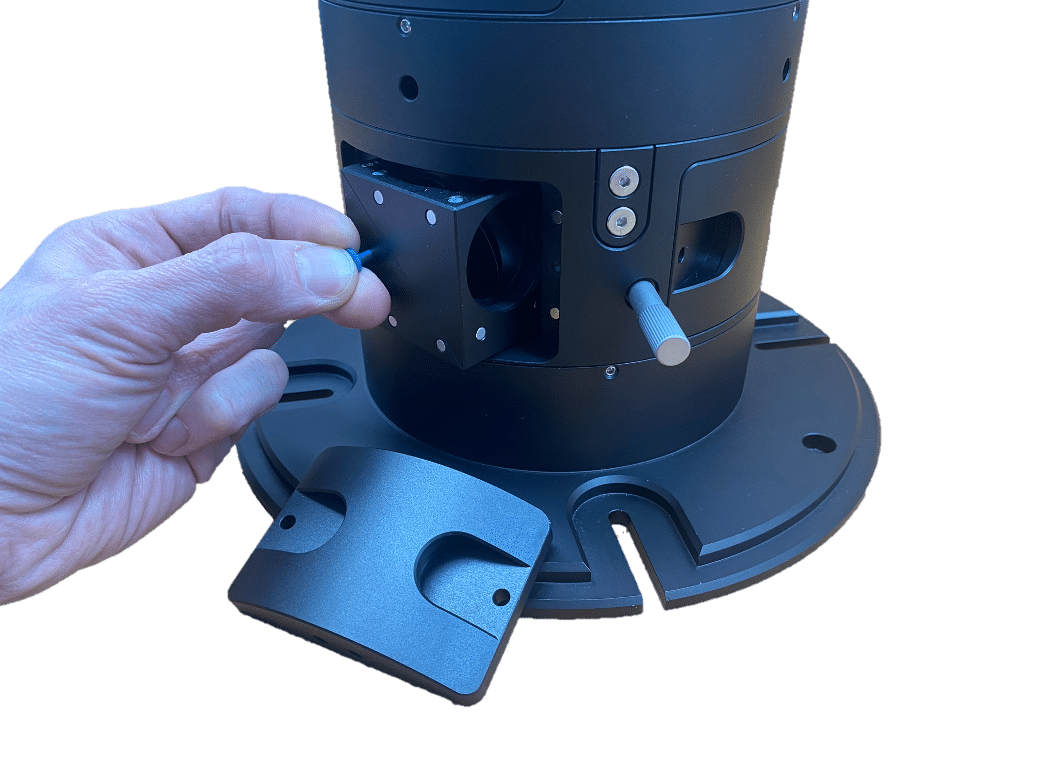
At the heart of the layer sits a filter cube that typically holds the dichroic mirror used to couple the illumination light into the central optical path of the openFrame. This filter cube is tip-tilt adjustable, to allow for precise alignment of the illumination. This is easiest achieved by viewing the illumination live on the camera and adjusting the resulting image to a target image


When used for Detection
On the openFrame the Single Detection Layer provides the means of connecting a single camera or similar detector to the microscope. This is the preferred solution for all applications requiring a single camera. It is also the recommended solution for systems that require simultaneous detection on multiple cameras. Here, the Flexi Layer would be populated with the first detection module and a beam splitter..
When using the Single Detection Layer, there are two possible camera mounting points located on opposing sides of the module. While only one of these is used, the two options provide more flexibility and avoid spatial conflicts with components on other layers, when bulky components are used.
At the heart of the layer sits a filter cube that typically holds a dichroic mirror and emission filter. The cube is used to reflect the desired fluorescence wavelength to the camera. This filter cube can easily be tip-tilt adjusted, to allow for pixel alignment of the camera. This is easiest achieved by viewing the live image on the camera and adjusting it to a target image.


Filter Cubes


